
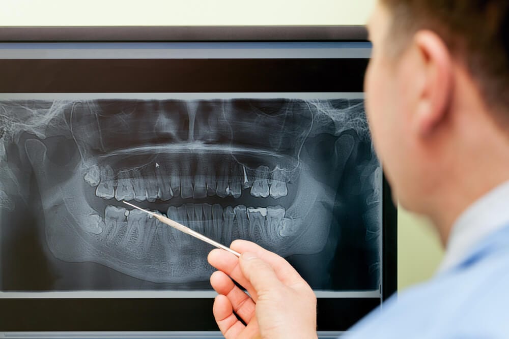
Digital X-rays are an important diagnostic tool for dental professionals. While traditional film radiographs provide critical insight into the oral and physical health of the patient, hi-tech digital radiographs allow dentists to view and enhance dental images on a large computer screen.
Dentists can also copy or print digital radiographs with ease. This allows for effective comparison of new results to previous images and insight on how treatments have impacted dental conditions. If the dentist refers the patient to a specialist, digital radiographs can be transmitted via computer – eliminating the need for a second set of x-rays in most cases.
Why Use Digital X-Rays?
One of the most significant advantages of utilizing digital radiographs is reduction of radiation exposure. Digital radiographs also eliminate the use of film and required chemicals for processing, making the overall procedure much less harmful to the environment.
The larger computer screen used to display digital radiographs allows dentists to view any problems or irregularities with added clarity. The potential for early detection of decay or periodontal problems and reducing complicated conditions later is vastly increased.
Here are some of the main conditions that digital radiographs can better expose:
- Small areas of decay
- Bone recession
- Tumors
- Fractures and trauma
- Positioning of the teeth
- Developmental irregularities
- Tooth positioning
How Are Digital X-Rays Taken?
The technique for capturing digital radiographs is similar to that of the traditional-style radiographs, but the digital variety uses a small electronic sensor to capture intraoral images, as opposed to film bitewings.
Generally, a full mouth series of digital x-rays includes eighteen different views of the teeth and underlying jawbone. The two standard views dentists use are: periapical and bitewing. The periapical view is used to inspect the root tips for decay, disease or damage, while the bitewing view allows for close inspection and measurement of the mandible and maxilla (upper and lower jawbones).
After exposure, the digital image is either transferred wirelessly to a computer, or the dentist takes the plate from the mouth, and scans it with a specialized reader. Processing traditional film can take up to five minutes, but a digital image takes mere seconds. Once the image is apparent on the screen, the contrast, color and brightness can be altered to produce a much clearer image.
If you have questions or concerns about getting a digital radiograph, please contact your dentist.
Are dental X-rays safe?
We are all exposed to natural radiation in our environment. The amount of radiation exposure from a full mouth series of X-rays is equal to the amount a person receives in a single day from natural sources.
Dental X-rays produce a low level of radiation and are considered safe. Dentists take necessary precautions to limit the patient’s exposure to radiation when taking dental X-rays. These precautions include using lead apron shields to protect the body and using modern, fast film that cuts down the exposure time of each X-ray.
How often should dental X-rays be taken?
The need for dental X-rays depends on each patient’s individual dental health needs. Your dentist and dental hygienist will recommend necessary x-rays based on the review of your medical and dental history, dental exam, signs and symptoms, age consideration, and risk for disease.
A full mouth series of dental X-rays is recommended for new patients. A full series is usually good for three to five years. Bite-wing X-rays (X-rays of top and bottom teeth biting together) are taken at recall (check-up) visits and are recommended once or twice a year to detect new dental problems.
PANORAMIC X-RAYS
Panoramic X-rays (also known as Panorex® or orthopantomograms) are wraparound photographs of the face and teeth. They offer a view that would otherwise be invisible to the naked eye. X-rays in general, expose hidden structures, such as wisdom teeth, reveal preliminary signs of cavities, and also show fractures and bone loss.Panoramic X-rays are extraoral and simple to perform. Usually, dental X-rays involve the film being placed inside the mouth, but panoramic film is hidden inside a mechanism that rotates around the outside of the head.Unlike bitewing X-rays that need to be taken every few years, panoramic X-rays are generally only taken on an as-needed basis. A panoramic X-ray is not conducted to give a detailed view of each tooth, but rather to provide a better view of the sinus areas, nasal areas and mandibular nerve. Panoramic X-rays are preferable to bitewing X-rays when a patient is in extreme pain, and when a sinus problem is suspected to have caused dental problems.Panoramic X-rays are extremely versatile in dentistry, and are used to:Assess patients with an extreme gag reflex.Evaluate the progression of TMJ.Expose cysts and abnormalities.Expose impacted teeth.Expose jawbone fractures.Plan treatment (full and partial dentures, braces and implants).Reveal gum disease and cavities.How are panoramic X-rays taken?The panoramic X-ray provides the dentist with an ear-to-ear two-dimensional view of both the upper and lower jaw. The most common uses for panoramic X-rays are to reveal the positioning of wisdom teeth and to check whether dental implants will affect the mandibular nerve (the nerve extending toward the lower lip).The Panorex equipment consists of a rotating arm that holds the X-ray generator, and a moving film attachment that holds the pictures. The head is positioned between these two devices. The X-ray generator moves around the head taking pictures as orthogonally as possible. The positioning of the head and body is what determines how sharp, clear and useful the X-rays will be to the dentist. The pictures are magnified by as much as 30% to ensure that even the minutest detail will be noted.Panoramic X-rays are an important diagnostic tool and are also valuable for planning future treatment. They are safer than other types of X-rays because less radiation enters the body.If you have questions or concerns about panoramic X-rays, please contact our practice.
CEPHALOMETRIC X-RAYS
The cephalometric X-ray is a unique tool that enables the dentist to capture a complete radiographic image of the side of the face. X-rays in general offer the dentist a way to view the teeth, jawbone, and soft tissues beyond what can be seen with the naked eye. Cephalometric X-rays are extraoral, meaning that no plates or film are inserted inside the mouth. Cephalometric and panoramic X-rays display the nasal and sinus passages, which are missed by intraoral bitewing X-rays.
Cephalometric X-rays are usually taken with a panoramic X-ray machine. The adapted machine will have a special cephalometric film holder mounted on a mechanical arm. An X-ray image receptor is exposed to ionizing radiation in order to provide the dentist with pictures of the entire oral structure. The advantage of both cephalometric and panoramic X-rays is that the body is exposed to less radiation.
Cephalometric X-rays are not as common as “full sets” or bitewing X-rays, but they serve several important functions:
- Provide views of the side profile of the face.
- Provide views of the jaw in relation to the cheekbone.
- Provide information about “bad bites” or malocclusions.
- Allow measurement of the teeth.
- Identify fractures and other injuries to the teeth and jawbone.
- Assists in orthodontic planning.
How are cephalometric X-rays taken?
Cephalometric X-rays are completely painless. The head is placed between the mechanical rotating arm and the film holder, which is placed on another arm. The arm rotates around the head capturing images of the face, mouth, and teeth. The clarity and sharpness of these images will depend on the positioning of the body. The images are usually magnified up to 30%, so any signs of decay, disease, or injury can be seen and treated.
After capturing cephalometric X-rays, the dentist will be able to see a complete side profile of the head. This can assist in orthodontic planning and allow an immediate evaluation of how braces might impact the facial profile and teeth. Another common use for this type of X-ray is to determine specific measurements prior to the creation and placement of dental implants.
If you have any questions or concerns about cephalometric X-rays, please ask our dentist.